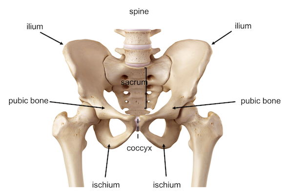
In order to understand the pathology trochanteric or hip bursitis, let’s begin with some anatomical descriptions. The hip is one of the most complex joints of the body. It consists of a ball-and-socket joint connecting the femoral head to the acetabulum at the pelvis. The femoral head continues distally with the femoral neck and the greater trochanter. The latter is the protruding portion of the femur that can be felt laterally at the upper thigh, below the crest of the pelvis.

Anatomy of the pelvis and hip showing the femoral head inserting into the acetabulum at the pelvis to form the hip joint
The hip joint is protected by articular cartilage, which is a layer of glassy yet tough connective tissue surrounding both the femoral head and the acetabulum. The cartilage facilitates the movement of the joint by reducing the friction of the femoral head rotating inside the socket and importantly it absorbs shock impacts. Overuse of the cartilage often leads to osteoarthritis of the hip due to the excessive, frequent load to the hip joint with physical activity.

Anatomy of the femur including the greater trochanter where the bursa is located
The hip joint allows a wide range of movements of the lower limbs and is used when walking, running, climbing, lunging and bending. Because it bears the body weight, it is constantly under stress and thus supported by large muscles, strong tendons and ligaments.
The hip bursas
The hip joint is also supported by the bursas. A bursa is a fluid-filled sac, functioning as a cushion to absorb shock and facilitate the gliding of muscles and bones around the joint. The hip has two main bursas:

There are a few bursas around the hip joint with the main ones being the trochanteric and ischio-gluteal bursa more often affected by the condition bursitis
The trochanteric bursa is located on the great trochanter where the gluteus maximus muscle of the hip joint is attached. This is quite a large bursa and is known for the related pathology, hip bursitis or trochanteric bursitis.
The ilio-psoas bursa is located on the inner side of the hip where the ilio-psoas muscle attaches to the lesser trochanter. Also, this bursa is subject to inflammation or bursitis, albeit less commonly.
Trochanteric bursitis
Trochanteric bursitis is an inflammatory condition of the bursa causing swelling and strong pain to the lateral side of the hip. It is a frequently occurring pathology originating from a number of reasons such as trauma, overuse, incorrect posture, leg length discrepancy, overweight, stress of the surrounding soft tissues, surgery, formation of hip spurs (bony growths) and other conditions such as rheumatoid arthritis, gout, psoriasis, thyroid disease and rarely, from infection.

Trochanteric bursitis is usually treated conservatively with anti-inflammatory therapy, physiotherapy and local injection of steroids and more recently also with platelet-rich-plasma (PrP). However, at times the bursa needs to be removed surgically either by open surgery or arthroscopy. Once diagnosed, trochanteric bursitis requires addressing changes in physical activities, posture and sporting techniques so that a re-injury is avoided.
If you wish to learn more on the issues relating to this Pathology, follow the link
For the Anatomy of the pelvis and hip click here
For the Examination of the pelvis and hip click here
The content and images found in any educational material released by Lex Medicus and Lex Medicus Publishing are protected by copyright.