The instability of the shoulder occurs when the tissues surrounding the shoulder joint are too loose, thus increasing the risk of the humeral head slipping out of the joint socket and dislocating.
But which joint of the shoulder is involved in this pathology? Why does it happen, and what are the causes, risk factors and associated pathologies? In order to understand, let’s begin with a few simple anatomical references to fully visualise the structures leading to shoulder instability.
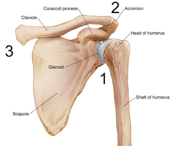
Shoulder anatomy
The shoulder is one of the most complex joints in the human body having almost a 360 degree range of motion. It is actually formed by three joints that are described below. This extensive mobility allows for the multiple actions of the arms and hands. For this reason, it must be stable enough to execute lifting, pushing and pulling. The compromise between mobility and stability may result in a number of shoulder conditions.
Bones of the shoulder
The shoulder consists of the head of the humerus fitting in to the glenoid fossa, a depression of the scapula (shoulder blade). These two parts fit with each other to form a socket ball joint. These bones are supported in the anterior part of the body by the clavicle, or collarbone, and in the posterior side by the acromion, the roof of the scapula. The cartilage covering the shoulder joint confers structural support to reduce friction of the articulating elements and facilitate a wide range of movements.

Shoulder joints
The movement of the shoulder is permitted by three distinct joints as illustrated above:
1. glenohumeral joint is the main joint of the shoulder consisting of the humeral head inserting in the glenoid
2. acromioclavicular (AC) joint formed by the acromion, which is the postero-superior portion of the scapula and the clavicle
3. sternoclavicular (SC) joint situated at the centre of the upper chest right under the throat where both clavicle bones meet the sternum (breast bone)
The shoulder capsule
To keep the head of the humerus within the glenoid, a robust yet flexible connective tissue forms the shoulder capsule, which is the major ligament system of the shoulder. The capsule is formed by the glenohumeral ligaments and encloses the entire shoulder joint. It attaches the upper part of the humerus to the shoulder blade.

The glenoid labrum is a fibro-cartilaginous elastic structure that encloses the glenoid cavity creating a deep socket where the glenohumeral joint rests. At the superior side is the attachment to the long head of the biceps brachii muscle. The glenoid labrum is essential to providing stability to the shoulder joint.
Rotator cuff
The ligaments’ pivotal role is to connect bones to other bones, whereas tendons connect muscles to bones. The rotator cuff is an assembly of tendons originating from the muscles subscapularis, on the anterior side, and the supraspinatus, infraspinatus and teres minor (on the posterior side) joined together to form a cuff around the shoulder.

Legend: Left anterior view of the shoulder; Right, posterior view of the shoulder
What is the cause of shoulder instability?
The instability of the shoulder is a chronic condition resulting from the laxity of the glenohumeral joint that increases the risk of dislocations. This pathology can affect one or both sides. Joint laxity is caused by excessive stretching, micro-injuries and overuse of the shoulder ligaments that form the rotator cuff and the labrum. Consequently, the ligaments of the shoulder are unable to keep the humerus head within the socket, thus facilitating the subluxation or full dislocation when performing overhead movements. Shoulder instability can lead to tears of the ligaments and other structures (Bankart lesion) and cause severe osteoarthritis of the shoulder joint.
Frequent shoulder subluxations can also stretch and even damage the axillary nerve extending below the humeral head. The pathology is often observed in athletes performing frequent throwing/overhead action (cricket, swimming) or in those with congenital connective tissue laxity (Ehler Danlos Syndrome).
Symptoms and medico-legal examination
The sensation of shoulder instability is felt when the arm is raised in shoulder flexion triggering pain. If a dislocation occurs the contour of the shoulder is clearly altered and require the reposition of the humeral head in the glenoid socket with or without anaesthesia.

During the medico-legal examination, the specialist will perform the apprehension test by passively externally rotating the patient’s arm in 90° abduction and applying forward pressure to the posterior side of the humeral head. The test is positive when the patient reports pain.
The treatment for shoulder instability is usually conservative aiming at reducing the pain with anti-inflammatory drugs and later on strengthening the muscles forming the rotator cuff. Surgical intervention is recommended in more complicated cases with two procedures named capsular shift and thermal capsule shrinkage.
If you wish to know about these procedures and read the full pathology with additional details on definitions, causes, risk factors, treatments and prevention, click this link.
Disclaimer: All images and content published in the Lex Medicus, and Lex Medicus Publishing websites are protected by copyright.