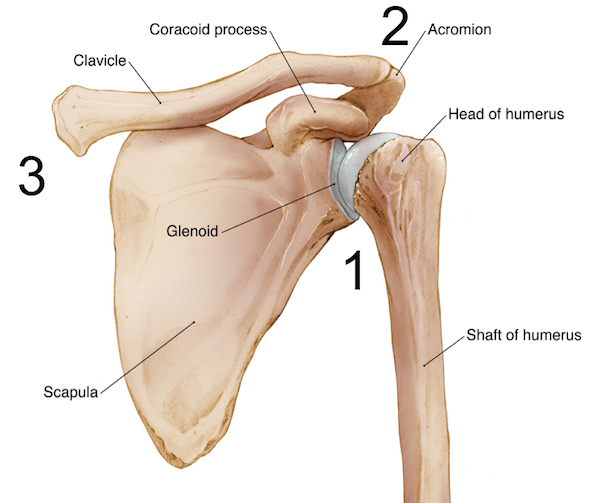
Bankart and Hill-Sachs lesions are injuries involving the gleno-humeral joint of the shoulder following one or multiple shoulder dislocations. These injuries affect the glenoid fossa on the scapular side (Bankart lesion) but also damage to the head of the humerus (Hill-Sachs lesion).
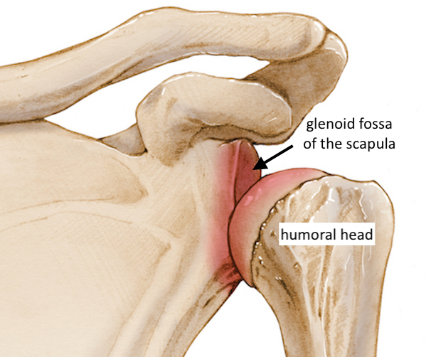
Bankart lesion
Two different types of Bankart lesions have been described, one with and one without the fracture of the glenoid.
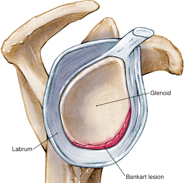
A Bankart lesion without a fracture of the glenoid is in most cases a lesion of the glenoid labrum (the glenoid rim; see anatomical definition below) caused by a shoulder dislocation and/or repeated anterior shoulder subluxations. With an anterior dislocation of the shoulder, the damage of the glenoid labrum can lead to the partial detachment of the labrum from the glenoid at its antero-inferior portion.
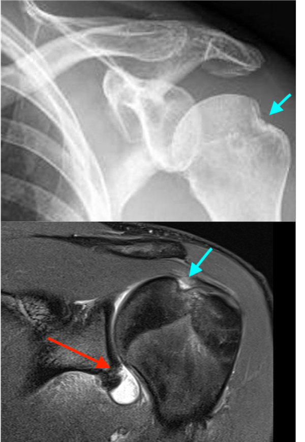
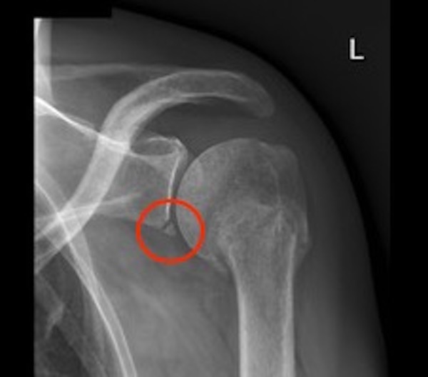
An osseous Bankart lesion occurs in patients with a full anterior shoulder dislocation, resulting in a more significant injury due to the detachment of the antero–inferior labrum associated with a glenoid rim fracture. Radiological images are critical for detecting an osseous Bankart lesion of glenoid bone and allow for the measurement of bone loss.
Hill-Sachs lesion
A Hill–Sachs lesion, or Hill–Sachs impaction fracture is an injury to the postero-lateral side of the humeral head. This injury is also caused by a shoulder dislocation. The name of this pathology derives from the American radiologists who first described it in 1940.
This humeral head lesion occurs mostly in young individuals and shows an incidence of 35% of all anterior dislocations and up to 80% in recurring dislocations.
Both pathologies (Bankart and Hill-Sachs lesions) can be associated with axillary nerve injury and larger fractures of the humeral head and humerus bone.
Anatomy of the shoulder
To fully comprehend this pathology affecting the gleno-humeral joint, it is important to define the main anatomical structures of the shoulder, its multiple bones and joints. Together they allow an almost 360 degree range of movement, the widest of any socket-ball joints of our body.
Three bones
The shoulder consists of the head of the humerus fitting in to the glenoid fossa, a depression of the scapula (or shoulder blade). These two parts fit with each other to form a socket-ball joint.
The humerus and the scapula are supported anteriorly by the clavicle, or collarbone, and posteriorly by the acromion, the roof of the scapula. The cartilage covering the shoulder joints confers structural support to reduce friction of the articulating bones and facilitate a wide range of movements.
Three joints
The shoulder is formed by three distinct joints:
- The gleno-humeral joint which is the main joint of the shoulder consisting of the humeral head inserted and surrounded by the glenoid;
- The acromio-clavicular (AC) joint formed by the acromion, which is the postero-anterior bone of the scapula and the clavicle or collar bone;
- The sterno-clavicular (SC) joint situated at the sternum right under the throat.
The gleno-humeral joint is among all three shoulder joints the one that is more frequently affected by injuries such as dislocation and pathologies involving the connective structures that help keep the joint together. These include the ligaments of the shoulder capsule, the tendons forming the shoulder capsule and the subacromial/subdeltoid bursa.
What is the shoulder capsule?
To keep the head of the humerus within the glenoid, a robust and yet flexible connective tissue forms the shoulder capsule, which is the major ligament system of the shoulder. The capsule consists of the gleno-humeral ligaments and encloses the entire shoulder joint. It attaches the upper part of the humerus to the shoulder blade.

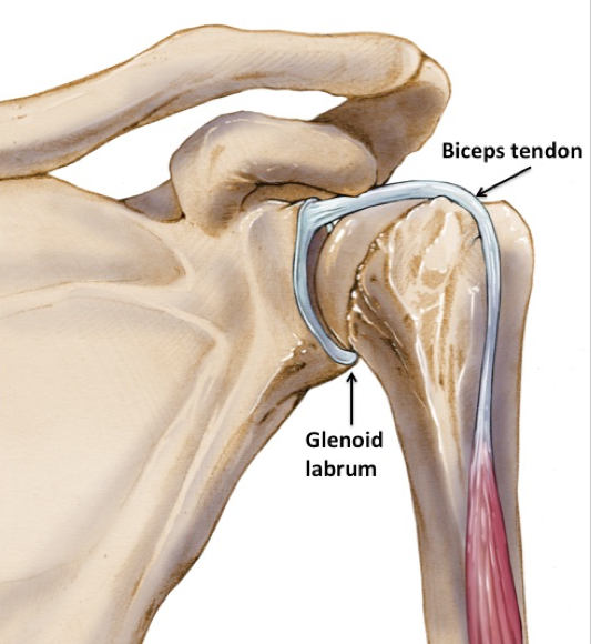
The glenoid labrum is a fibro-cartilaginous elastic structure that encloses the glenoid cavity creating a deep socket where the humeral head firmly rests. The glenoid labrum is essential to provide stability to the shoulder joint.
If you wish to know more about the causes, risk factors, symptoms, diagnosis, conservative, surgical and rehabilitative treatments, and prevention of Bankart & Hill-Sachs lesions please follow the link: https://pathologies.lexmedicus.com.au/collection/bankart-lesion-and-hill-sachs-lesion
For the shoulder anatomy: https://anatomy.lexmedicus.com.au/collection/shoulder
For the examination of the shoulder to understand changes in the range of movement as assessed by a medical examiner: https://examination.lexmedicus.com.au/collection/shoulder
