Before delving into today’s pathology, ulnar nerve entrapment, I would like to first introduce a few fun facts on the anatomy of the elbow and arm including the description of all three main nerves: the median, radial and ulnar nerve.
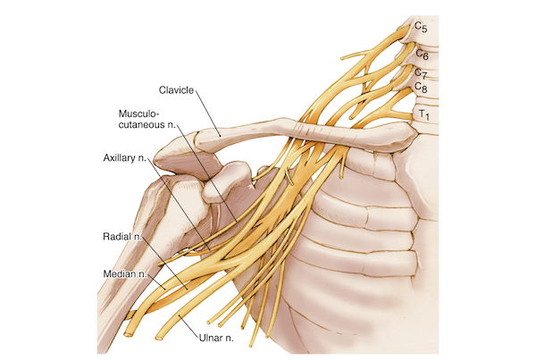
Like all nerves of the upper extremities, the ulnar nerve originates from the C8 and thoracic nerve roots of the cervical spine. From the neck, these nerves descend along the body to form the brachial plexus from where they extent along the shoulder, the upper arm and forearm down to the hand and fingers.
The main nerves of the shoulder / upper extremities are the axillary nerve, the median, the radial and the ulnar nerve.
Trauma and other causes can impact on these nerves and/or their soft connective tissue, consequently leading to pain, numbness and weakness in the arm and hand.
Anatomical features of the ulnar nerve
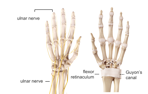
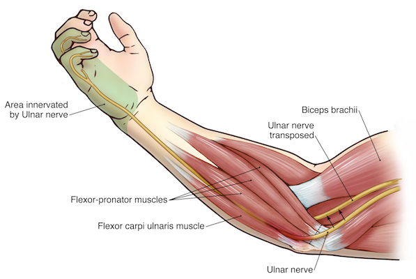
At the elbow, the ulnar nerve lies under the groove of medial epicondyle (inner side of the elbow at the distal extremity of the humerus), whereby connective tissues closes the groove. This takes the name of cubital tunnel. Further distally, at the wrist, the other tunnel of the ulnar nerve is called Guyon’s canal.
Motor function of the ulnar nerve
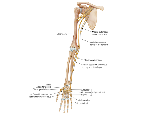
The motor branches of the ulnar nerve innervate the muscles of the forearm and hand allowing the flexion of the fingers to grip and exert fine hand movements; the ulnar nerve also contributes to the coordination of finger motion and pinch strength, as well as the flexion of the small and ring fingers.
Sensory function of the ulnar nerve
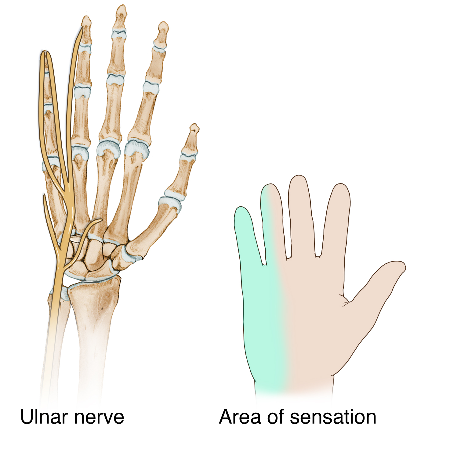
The ulnar nerve regulates the sensation to the little finger and the outer side of the ring finger (ulnar side) on both the palmar and dorsal sides of the hand. It also mediates parts of the sensation of the palm. This nerve is known for eliciting an electric shock type of reaction when hit at the elbow’s medial epicondyle.
The pathology
Ulnar nerve entrapment is caused by the swelling of the soft tissue surrounding the nerve, resulting in constriction of the nerve and functional neuropathy. This can occur at different levels along the nerve, such as the neck, collarbone and wrist, but it is mostly observed at the side of the elbow. In this case it is named cubital tunnel syndrome. If it occurs at the wrist it is called Guyon’s canal syndrome. Depending on the entrapment point, there are different neurological manifestations.
The aetiology of ulnar nerve entrapment at the elbow is unknown. Nerve compression can occur in association with:
– Degeneration of the cervical spine
– Fracture and dislocation to the elbow
– Prolonged leaning on the elbows
– Direct blow to the elbow at the “funny bone”
– Repetitive bending of the elbow
– Cyst in the elbow
Fracture and dislocation to the wrist can also cause compression at the Guyon’s canal.
Diagnosis
Tinel’s sign is the clinical test normally used to identify/diagnose ulnar nerve entrapment during physical examination. Further verification of ulnar nerve entrapment is performed by nerve conduction studies.

The examiner will tap on the medial side of the elbow and observes whether it triggers the symptoms. When the nerve is severely damaged the fingers may remain partially bent (ulnar claw). Hand clawing is more accentuated when the ulnar entrapment is located at the wrist (Guyon’s canal syndrome) but the sensation on the dorsal hand remains preserved.
Treatment
The treatment of ulnar nerve entrapment can vary from conservative, including the immobilisation of the elbow, steroid injection, to surgical treatment, which may involve cubital tunnel release or the transposition of the ulnar nerve to the anterior side of the elbow.
If you are interested in learning about this pathology in more detail, including the causes, risk factors, symptoms and diagnosis, as well as the different approaches of treatment and prevention, please follow this link.
Disclaimer: All images and content published in the Lex Medicus, and Lex Medicus Publishing websites are protected by copyright.
